- Get link
- X
- Other Apps
262021 A choroid plexus cyst is a cyst that can grow in the brain of a fetus during development. Choroid plexus cysts can also be found.
 Chromosomal Anomalies Radiology Key
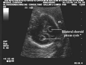
Unilateral choroid plexus cyst was seen.
Choroid plexus cyst ultrasound picture. 3232014 The following images were obtained. 1 article features images from this case Choroid plexus cyst antenatal. 3252019 Choroid Plexus Cysts CPC are small fluid filled areas in the brain and they are a common ultrasound finding in the fetus during the 2nd trimester of pregnancy.
Within a period of 25 years cystic structures in the choroid plexus were encountered at cerebral sonography in 70 neonates and babies 45 male 25 female. Their prevalence in patients examined during the first 4 weeks of life n 55 was 3. Anechoic lesions are noted within choroid plexuses.
The choroid plexus has the important function of producing cerebrospinal fluid. Essentially a soft marker in cerebral ventricles yet a frequent observation which should be mentioned and monitored is choroid plexus cyst. Ultrasound scan of a foetus brain with a choroid plexus cyst CPC.
No other abnormality was noted in rest of the fetal scan. A set of 20 fetal sonographic images with choroid plexus cysts were used to create cyst prototypes 1525 mm which were randomly embedded into normal choroid plexus images from varying gestational ages. Cysts are seen in the choroid plexus in about 1 out of every 50 or 100 pregnancies 1-2.
They are the most common type of intraventricular cyst. The number size and shape of the cysts can vary. It produces a fluid called cerebrospinal fluid that flows around the brain and provides a protective cushion for it against impacts.
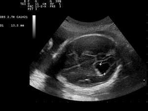
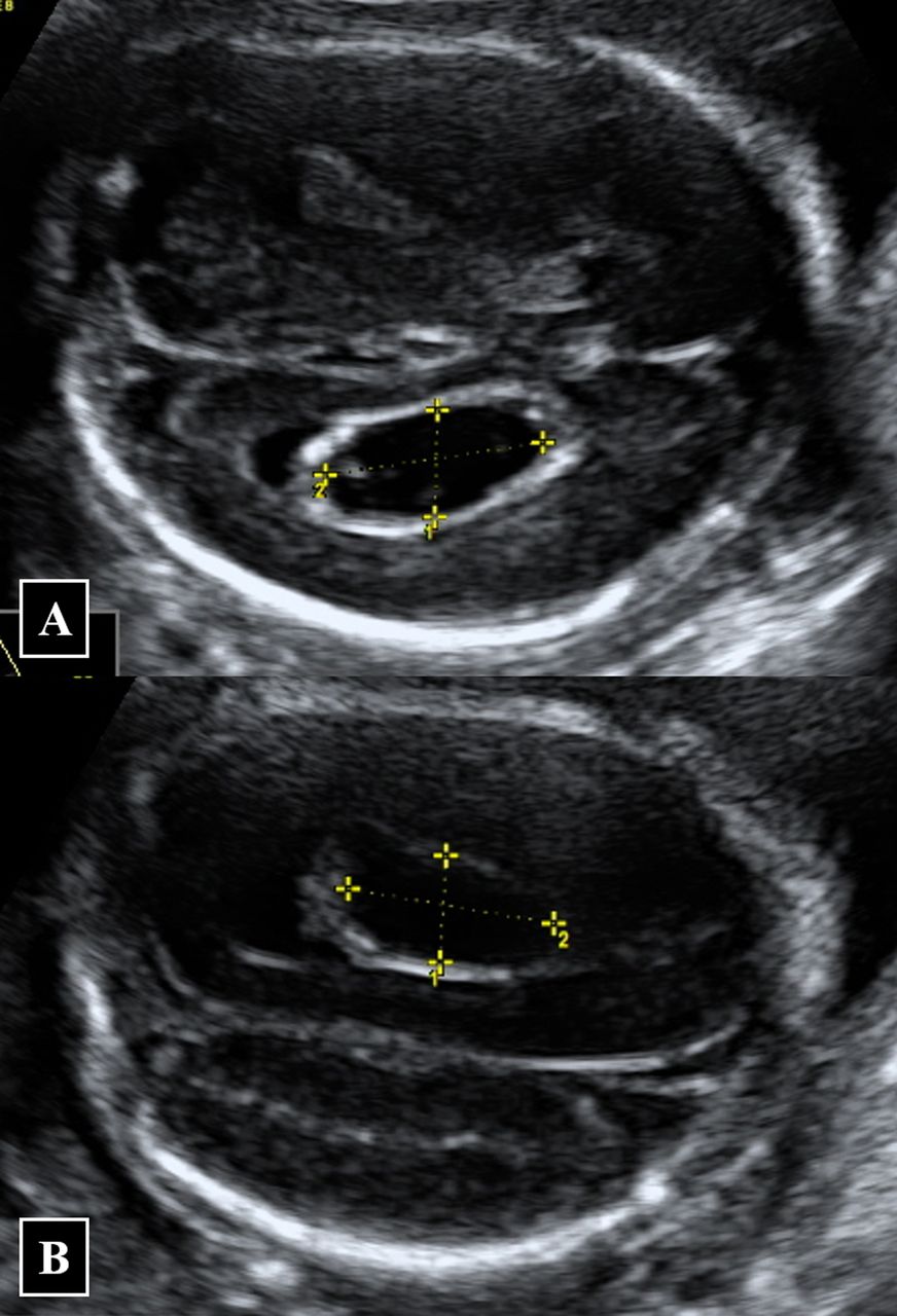
This is in the middle of the fetal brain. The fluid in the ventricles is produced by spongy tissue known as the choroid plexus that appears to float in. Right - 5 x 4 mm.
Located on the left and the right side of the brain the choroid plexus is a gland that produces cerebrospinal fluid. The brain contains pockets or spaces called ventricles with a spongy layer of cells and blood vessels called the choroid plexus. The appearance of a choroid plexus cyst generally is not a cause for alarm but these cysts sometimes indicate an increased risk of Down syndrome.
A normal finding on ultrasound. They are most commonly seen in the second and third trimester of the pregnancy and can be detected via an ultrasound. The fluid produced by the cells of the choroid plexus fills the ventricles and.
Choroid plexus cysts are seen at the same rate in fetuses with Down syndrome. Now for a short review of the choroid plexus cyst and its significance. A choroid cyst is a small buildup of fluid in the choroid plexus a structure in the brain which produces cerebrospinal fluid CSF.
Clinical significance of fetal choroid plexus cysts. This fluid bathes and protects the brain and spinal column. The exact cause of choroid plexus cyst is unknown.
How does it happen. How Common Are Choroid Plexus Cysts. 2152021 A choroid cyst can be found with help of an ultrasound.
In 1 to 2 of babies a cyst- a small round fluid filled area- is formed in the choroid plexus. In most instances these are a normal variant. A test set of 544 images was created which included 408 images with choroid plexus cysts and 136 images without choroid plexus cysts.
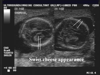
To identify a chroid plexus cyst CPC it must be imaged in two orthogonal planes and be greater than or equal to 3 mm in size. Choroid plexus cysts are seen during 1 to 3 of all second trimester ultrasound examinations. The choroid plexus is a layer of cells and blood vessels at the centre of the foetal brain.
Within each hemisphere there is a ventricle cavity or chamber that contains fluid that lines and protects the surrounding soft brain tissue. These glands make the fluid that normally circulates within the brain and spinal cord. They can be seen on one or both sides of the brain.
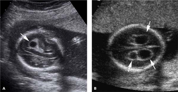
However choroid plexus cysts are seen much more frequently about 40 in fetuses with a condition called Edwards syndrome also known as trisomy 18. Left - 9 x 6 mm. The selected ultrasound images show small rounded well-defined cysts of the choroid plexus of the lateral ventricles measuring 7 mm on the right and 9 mm on the left.
Images of the other organs are given below. Choroid cysts are most commonly identified as an ultrasound finding and are in fact not uncommon being seen in around one to three percent of fetuses. Choroid plexus is not part of the brain involved in thinking or development.
Https Mydoctor Kaiserpermanente Org Ncal Images Gen Us 20cpc 20handout Tcm63 15061 Pdf
 Choroid Plexus Cyst Antenatal Radiology Reference Article Radiopaedia Org
 Choroid Plexus Cyst Radiology Case Radiopaedia Org
 Fetal Medicine Foundation Choroid Plexus Cyst Youtube
 Choroid Plexus Cyst Ultrasound Scan Stock Image M130 1007 Science Photo Library
 Medpix Case Trisomy 18
 Prenatal Ultrasound Findings A Choroid Plexus Cysts At 18 Weeks Of Download Scientific Diagram
 Choroid Plexus Anomalies Cysts And Papillomas Sciencedirect
Choroid Plexus Cyst Wikipedia
 Wk 6 L 2 Choroid Plexus Cyst Radiology Case Radiopaedia Org Diagnostic Medical Sonography Plexus Products Radiology
 Choroid Plexus Cyst Cpc Hkog Info
 Isolated Large Bilateral Choroid Plexus Cysts Associated With Trisomy 18 Bmj Case Reports
Comments
Post a Comment