- Get link
- X
- Other Apps
Our cardiologist and echo technicians do not use a contrast agent during this test. We hypothesised that the BS is frequently requested in patients that have a readily identifiable cause of stroke that any PFO detected is likely incidental and its detection often does not alter management.
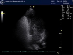
![]() Transthoracic Echocardiogram Bubble Study In A Supine Position And Download Scientific Diagram
Transthoracic Echocardiogram Bubble Study In A Supine Position And Download Scientific Diagram
Or transient ischemic attack.

Bubble study echocardiogram. The purpose of this study was to quantify the bubbles created by various quantities of agitated saline. Echocardiogram with bubble study. Sometimes the person doing an echocardiogram thinks the bubble study would be helpful and decides to do it then.
A non-invasive study typically done with an echocardiogram a bubble study helps your cardiologist to assess the blood flow and identify potential issues inside the heart. A bubble echocardiogram is typically. A bubble study is a noninvasive test that allows physicians to assess the flow of blood through the heart.
It can be especially helpful if someone has had a stroke or what is called a TIA. Once our policy is approved training complete and video reviewed please download the competency form and complete with one RN and the Cardiac Sonographer. This suspension was withdrawn 1 year later but an expanded list of contraindications to the use of Sonovue was inserted in the product.
Bubble contrast echocardiogram showed return of bubbles to left atrium between the third and fourth cardiac cycle suggestive of pulmonary arteriovenous shunt. The Bubble Study involves a small amount of micro bubbles being sent to the heart via a vein to watch how they travel through the heart. The echocardiographic BS is frequently performed in patients that have a readily identifiable cause of stroke and whose PFO unlikely relates to the strokeTIA.
The procedure is safe but the complication rate warrants informed. A saltwater solution called saline is mixed with a small amount of air to create tiny bubbles and then injected into your vein. Previous studies have recommended various safe amounts of agitated saline.
A bubble study is a type of echocardiogram which is the ultrasound of the heart. 1302020 What Exactly is Echocardiogram with Bubble Study. It is also known as Transcranial Doppler study or contrast echocardiography.
Bubble Study findings resulted in a change in management in the minority. 8262019 For the bubble study you will get an intravenous IV line in a vein in your arm. This fluid then circulates up to the right side of your heart and shows up on the echocardiogram image.
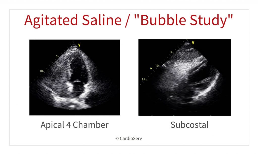
1212017 Such a study is labelled as negative contrast echocardiogram Fig. Bubble study Agitated saline transthoracic contrast echocardiograpyTTCE Contrast Echo Contrast Echocardiography. It is typically used in conjunction with an echocardiogram in which case doctors often call it contrast echocardiography or a transcranial Doppler study TCD.
Cardiac shunts are often identified using bubble studies in echocardiography with agitated saline. A doctor will discuss a bubble echocardiogram with a patient before its performed. Echocardiogram with Bubble Study Echocardiography is a scan that uses ultrasound sound waves to produce pictures of the heart.
During certain portions of the imaging saline with bubbles is injected into the vein. Other names for this test include. 1 and movie clip S1.
An echocardiogram is done to visualize the heart and its surrounding areas. Agitated Saline Echo Agitated Saline Echo what we order at Johns Hopkins Hospital to screen for PAVMs. Better images of the heart can be produced when a contrast is used during the echocardiogram.
812019 PFO is typically demonstrated with agitated saline bubble study BS during echocardiography. 6132009 In May 2004 the use of Sonovue for echocardiography was temporarily suspended in Europe by the EMEA 2 following 3 fatal cases in patients at high underlying risk for fatal cardiac complications which occurred after exposure to the agent. A bubble echocardiogram is the same procedure as an echocardiogram except an IV is placed in the patients arm.
ConclusionContrast echocardiogram is a sensitive tool to diagnose pulmonary arteriovenous shunt. Your doctor may ask to have a bubble study when the echocardiogram test is ordered. This poses a potential risk for air microembolism.
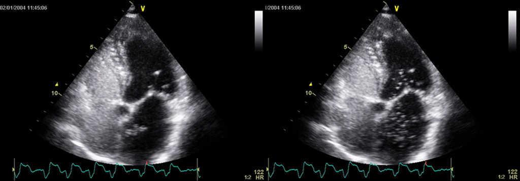
2142021 A bubble echocardiogram is a procedure which is designed to give a doctor an idea of how well someones heart is functioning. The appearance of these microbubbles in the left atrium LA left ventricle LV or aorta commonly referred to as positive contrast echocardiogram is diagnostic of a righttoleft shunt.
Anomalous Left Sided Superior Vena Cava With Cephalad Flow Ramani Gv Deible C Lopez Candales A Heart Views
 What Is A Bubble Study Harvard Health
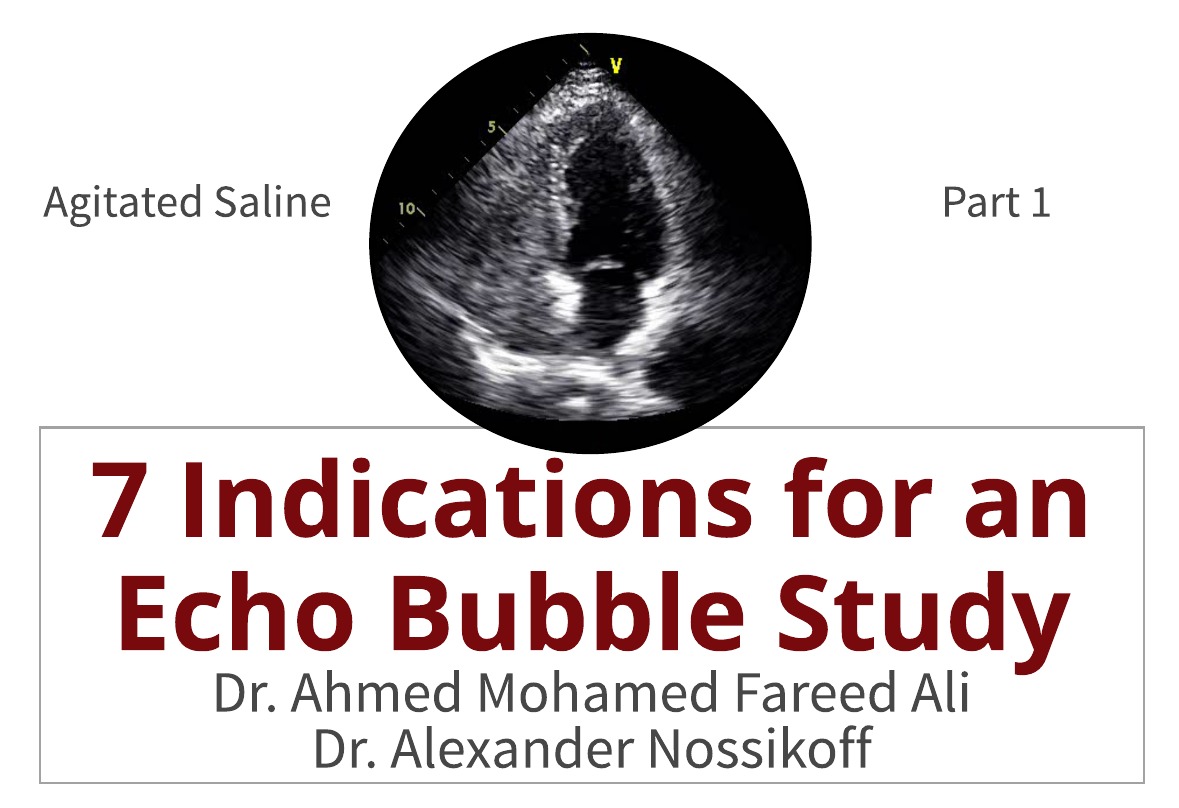 7 Indications For An Echo Bubble Study
 Echocardiogram With Bubble Study Youtube
 7 Indications For An Echo Bubble Study
 Andrew R Houghton How To Perform An Optimal Saline Bubble Contrast Echo Study
 Diagnosis And Quantification Of Patent Foramen Ovale Which Is The Reference Technique Simultaneous Study With Transcranial Doppler Transthoracic And Transesophageal Echocardiography Revista Espanola De Cardiologia
 A Bubble Study To Diagnose Patent Foramen Ovale Youtube
 Contrast Echo Thoracic Key
![]() Transesophageal Echocardiogram Tee Bubble Study Agitated Saline Download Scientific Diagram
Transesophageal Echocardiogram Tee Bubble Study Agitated Saline Download Scientific Diagram
 Bubble Contrast Echocardiography Bubble Echocardiogram Test Llc
![]() Transesophageal Echocardiogram Bubble Study A Even Though The Right Download Scientific Diagram
Transesophageal Echocardiogram Bubble Study A Even Though The Right Download Scientific Diagram
Http Www Rimed Org Rimedicaljournal 2018 08 2018 08 37 Case Powers Pdf
 Saline Contrast Echocardiography In The Era Of Multimodality Imaging Importance Of Bubbling It Right Gupta 2015 Echocardiography Wiley Online Library
Comments
Post a Comment